Room temperature correlative imaging

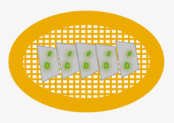
Fluorescence microscopy (FM) and electron microscopy (EM) are highly complementary techniques, and it can be beneficial to combine the information of both techniques from the same sample. This method is called correlative light- and electron microscopy (CLEM). Using fluorescence microscopy, regions of interest such as transfected cells, labelled proteins or rare events can easily be identified and mapped at relatively low magnification. The same regions of interest can then be targeted and examined at high resolution available in TEM. Most sample preparation methods for room temperature EM quench the fluorescence signal in a sample. Therefore, FM is typically done before EM sample preparation, and the same region of interest had to be found back in the EM.
An alternative RT-CLEM workflow is the fixation of samples by high-pressure freezing, followed by freeze-substitution, which allows FM and EM imaging of the section without any intermediate sample-preparations steps.
